Down Syndrome 3d Ultrasound Pictures
Certain findings sometimes called soft markers on.
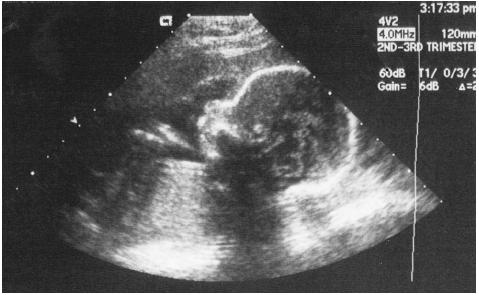
Down syndrome 3d ultrasound pictures. The detection of downs syndrome requires a sample of fetal cells from the fluid surrounding the fetus. C position positive in a fetus with down syndrome at 28 2 weeks. And d position negative in a trisomy18 fetus at 23. About 6000 babies are born with down syndrome each year in the united states or about 1 in every 700 births.
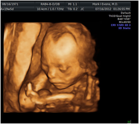
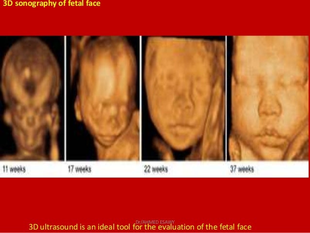
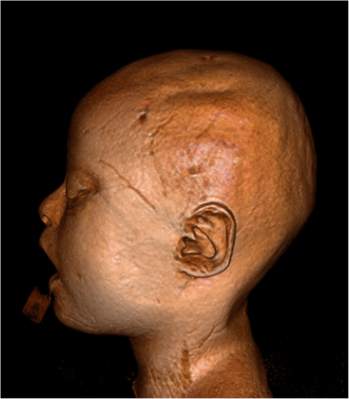
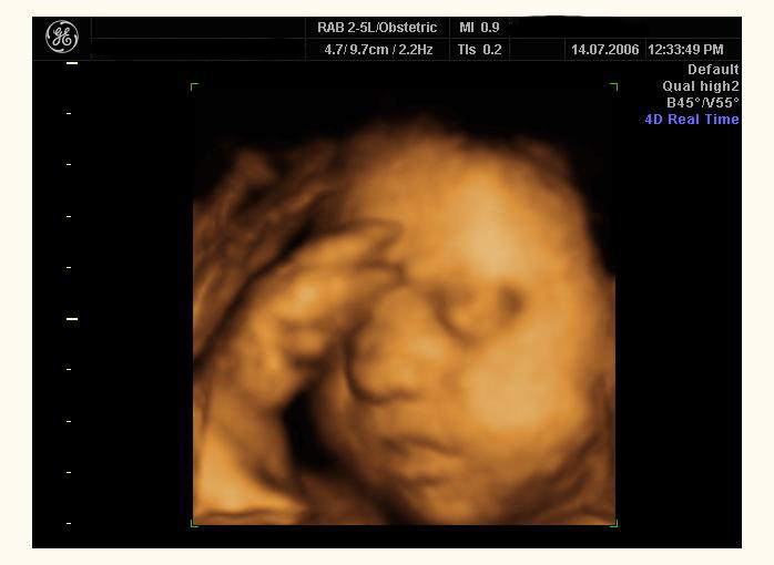
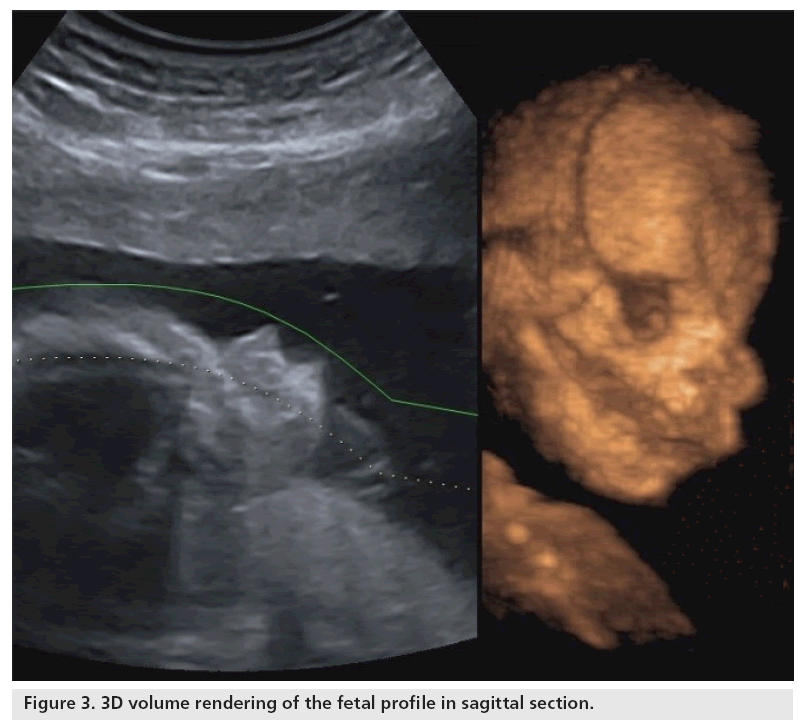
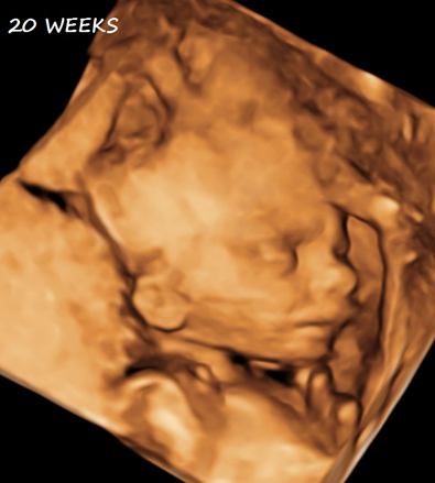
51 images obtained with 3d ultrasound in which the surface renderization enables us to obtain coronal oblique and sagittal planes of normal fetuses with gestational ages between 27 and 33 weeks 522 profile views in the profile view we study. B position zero in a fetus with down syndrome at 21 3 weeks. For this reason 3d ultrasound reconstruction of the nasal bone and other facial bones is useful. For example a fetus with down syndrome can have one nasal bone that appears normal and the second bone hypoplastic or absent.
A position zero in a euploid fetus at 24 6 weeks gestation. There are multiple prenatal genetic screening strategies and diagnostic tests aimed at accurate prenatal identification of down syndrome and other aneuploidies. Ultrasound cannot diagnose a fetus with down syndrome trisomy 21. Ultrasound in obstetrics and gynecology meta analysis of second trimester markers for trisomy 21 january 2013.
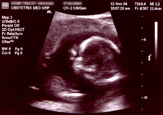
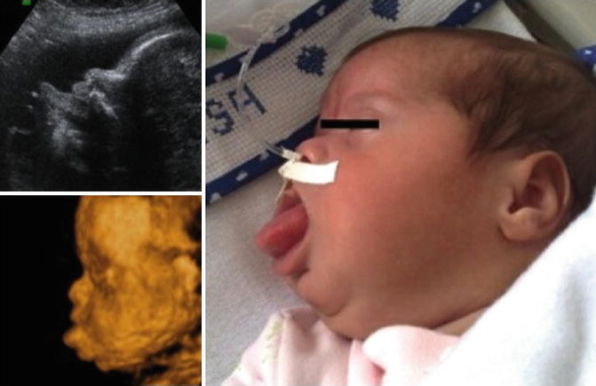
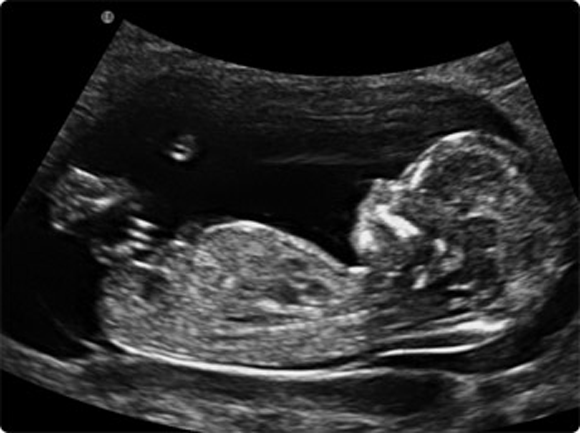
Modern ultrasound machines can detect most anomalies in the fetus including down syndrome and other chormosomal anomalies and other malformations. However they are seen more frequently in fetuses with an abnormality. If the 2d ultrasound does not demonstrate two nasal bones then 3d ultasound may be useful. Down syndrome trisomy 21 is the most common chromosomal disorder in live born infants.
However ultrasound is often used as a screening test for down syndrome and other chromosome abnormalities. Many but not all fetuses with down syndrome have one or more so called markers on ultrasound. Ultrasound in obstetrics and gynecology isolated fetal pyelectasis and the risk of down syndrome. The ultrasound examination cannot diagnose a fetus with down syndrome with certainty.
A meta analysis december 2013. It can pick up soft markers for downs. The forehead elusive or convex and discard prefrontal edema fig. Soft markers are sonographic findings that do not in themselves cause any adverse outcomes.
This test called an amniocentesis causes miscarriage in about 1 of women who have it. Approximately 30 of babies with down syndrome have detectable abnormalities on the mid trimester ultrasound 1.

Part 3 20 Week Scan Embracing Wade

Sonogram Secrets By Trimester Advanced Ultrasound Servicesadvanced Ultrasound Services

Ultrasound Nt Down Syndrome Test 12 Weeks Pregnant Down Syndrome Ultrasound Down Syndrome Test 12 Weeks Pregnant

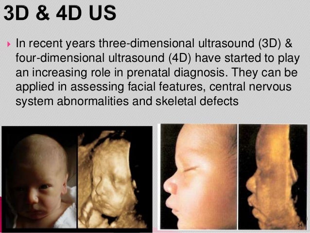
Ultrasound 3d And 4d What Is It What S The Difference Health Care Qsota

27 Weeks In The Womb Same Position Same Mad Face Perfect At Goldenview Ultrasound 3d Ultrasound 4d Ultrasound Baby Ultrasound


/babyboyultrasound-7bf2ced4b4794754b67dea974b7ec744.jpg)