When To Get 3d Ultrasound Done
This test creates images of the prostate by placing a special transducer into the rectum.

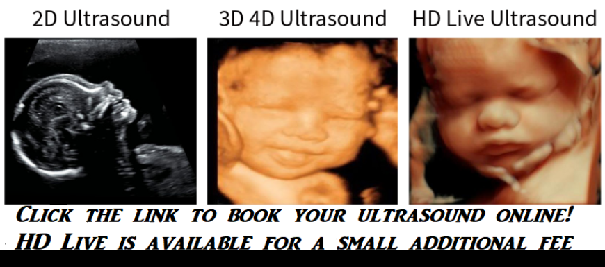
When to get 3d ultrasound done. Currently acog recommends that expecting women have at least one 2d ultrasound between weeks 18 to 22 of pregnancy noting that some women may also have a first trimester ultrasound. 3d ultrasound is done using sound waves which helps in capturing clear images of the uterus and the fetus for further study of the pregnancy. Its usually done while you are sedated. A second ultrasound or third ultrasound is usually done at 18 to 20 weeks to review the babys anatomy and rule out abnormalities.
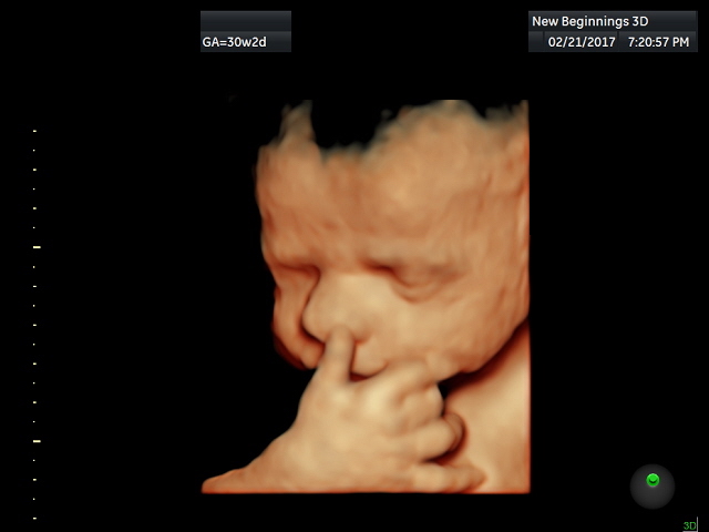
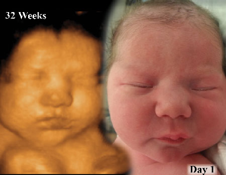
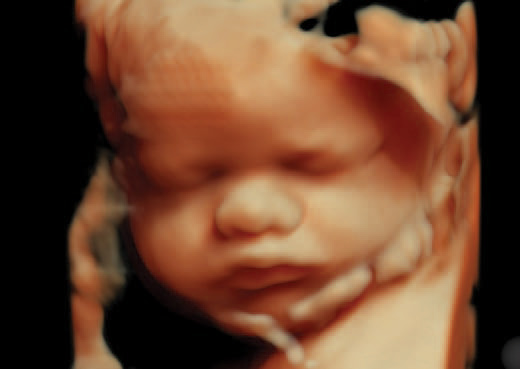
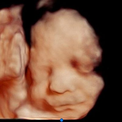
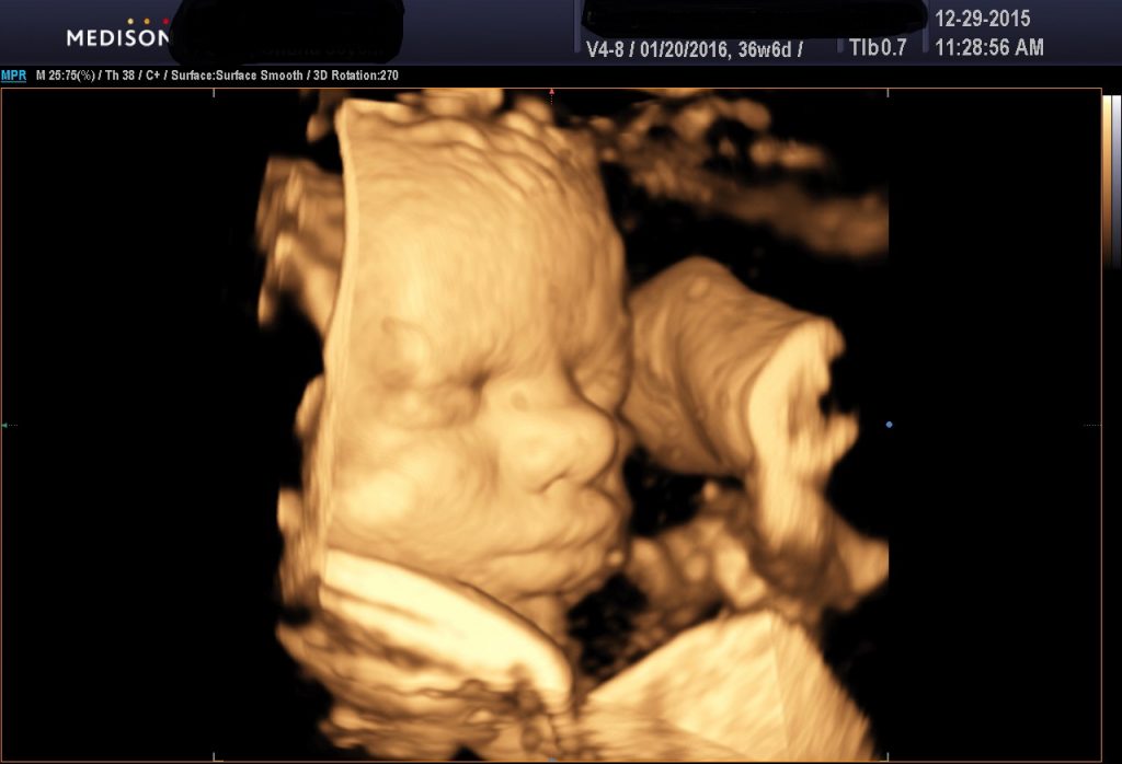
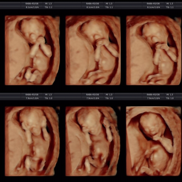
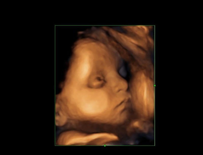
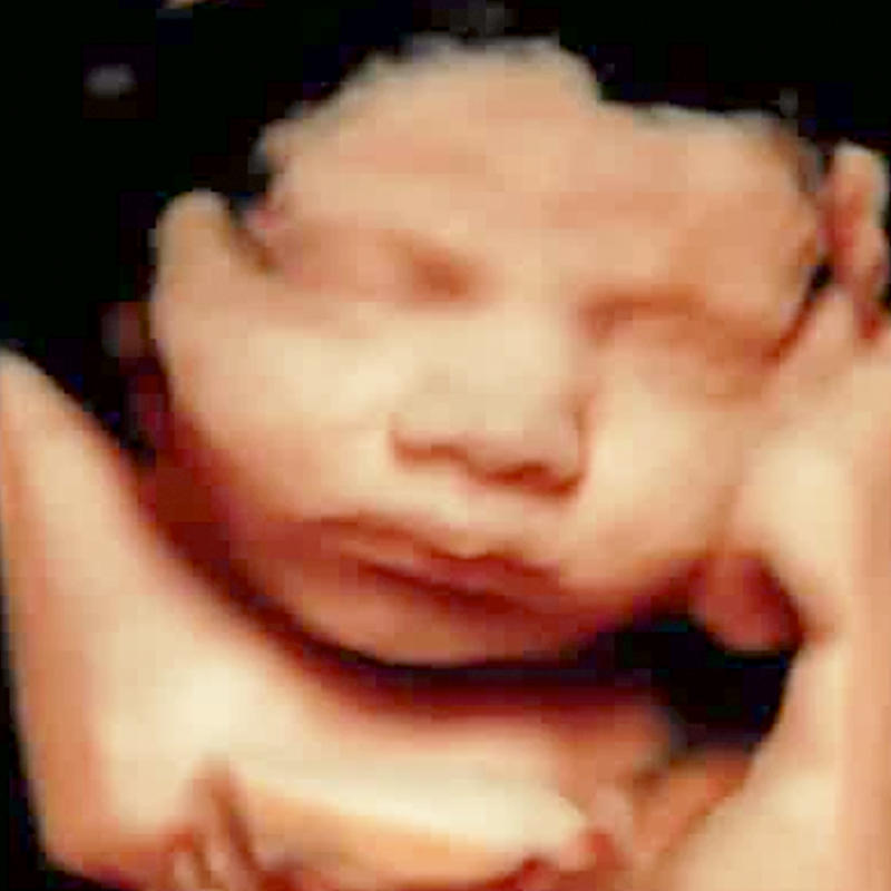
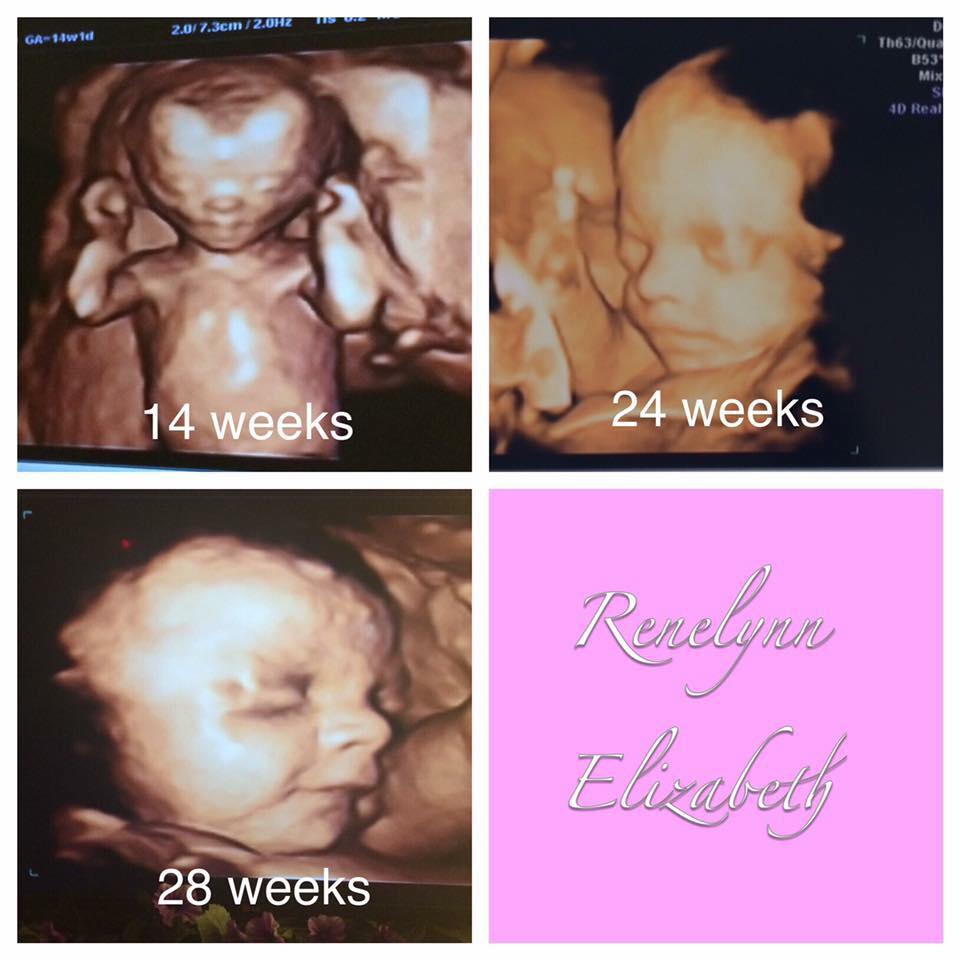
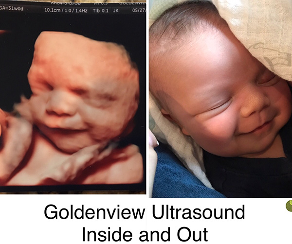
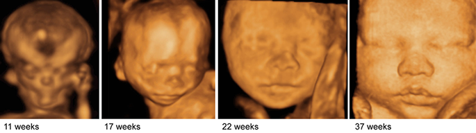
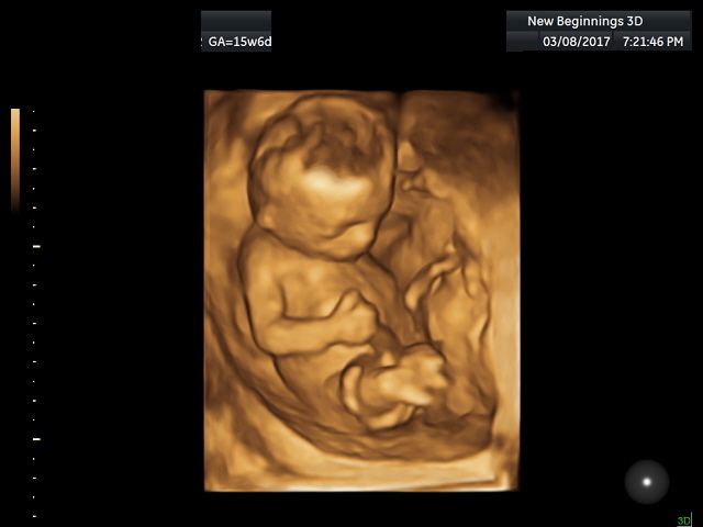
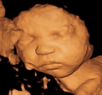
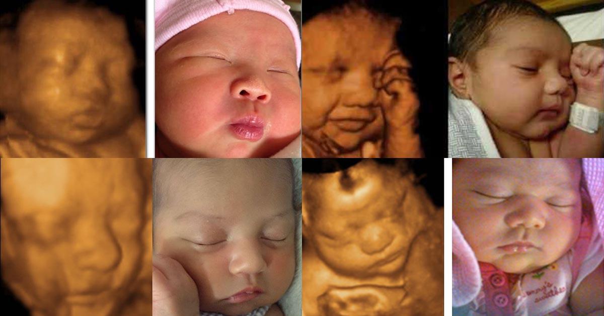
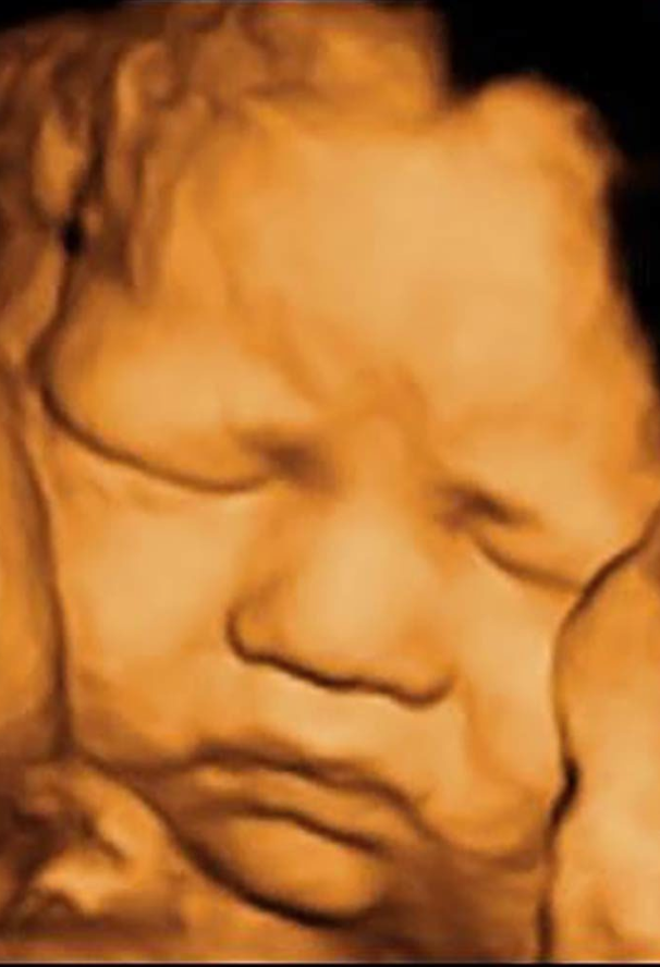
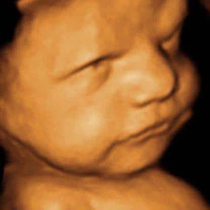
This may be done separately or at the same time as a scan offered at 11 13 weeks as part of the optional screening for trisomy 21 or down syndrome. To get good pictures 3d ultrasounds are best performed between 24 and 34 weeks and the best pictures are between 27 and 32 weeks. The best time to come in for a 3d4d ultrasound really depends on what you want to see. The 3d ultrasound is generally considered safe as there is no use of radiation insertion of any chemical solutions or use of needles and injections.
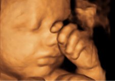
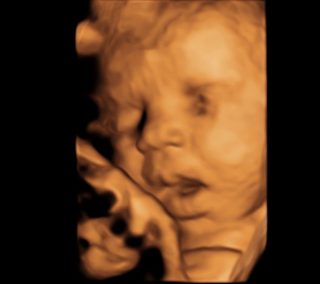
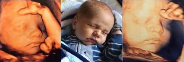
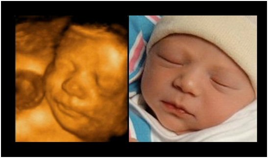
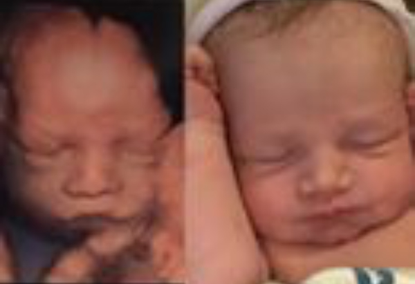
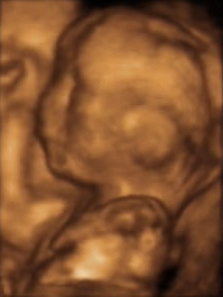
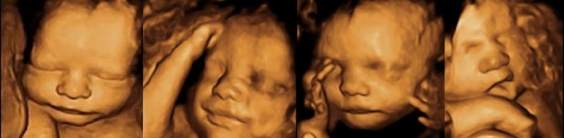
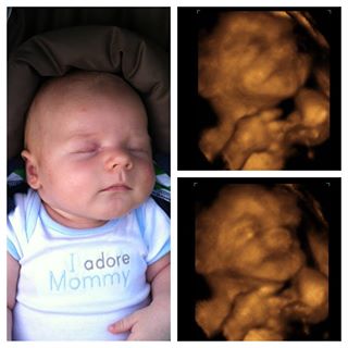
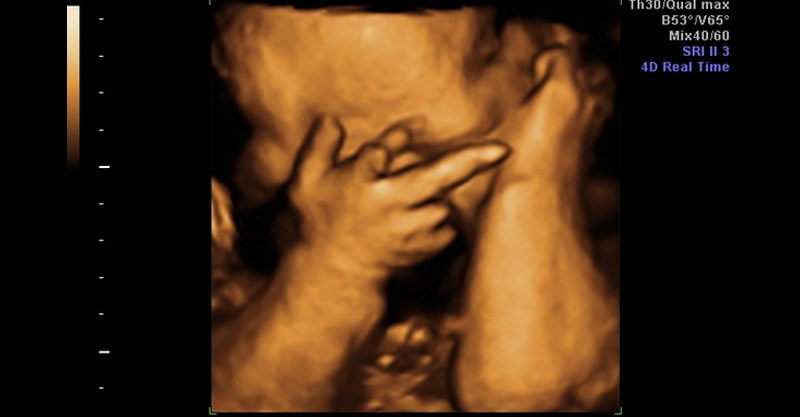
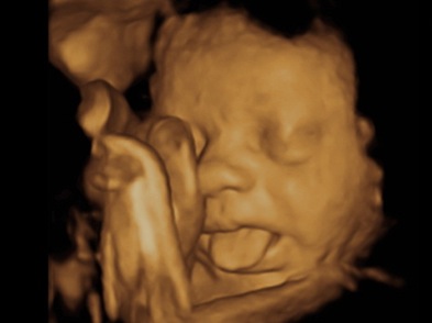
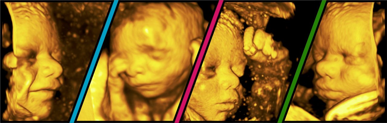
In fact many of these images also capture interesting activities in which the unborn baby is involved inside the womb. Also getting an ultrasound at a commercial center is not a substitute for medical care. Heshe might be seen sucking the thumb or playing etc. If you would like to get a closeup shot of the face then 27 to 28 weeks is the best time.
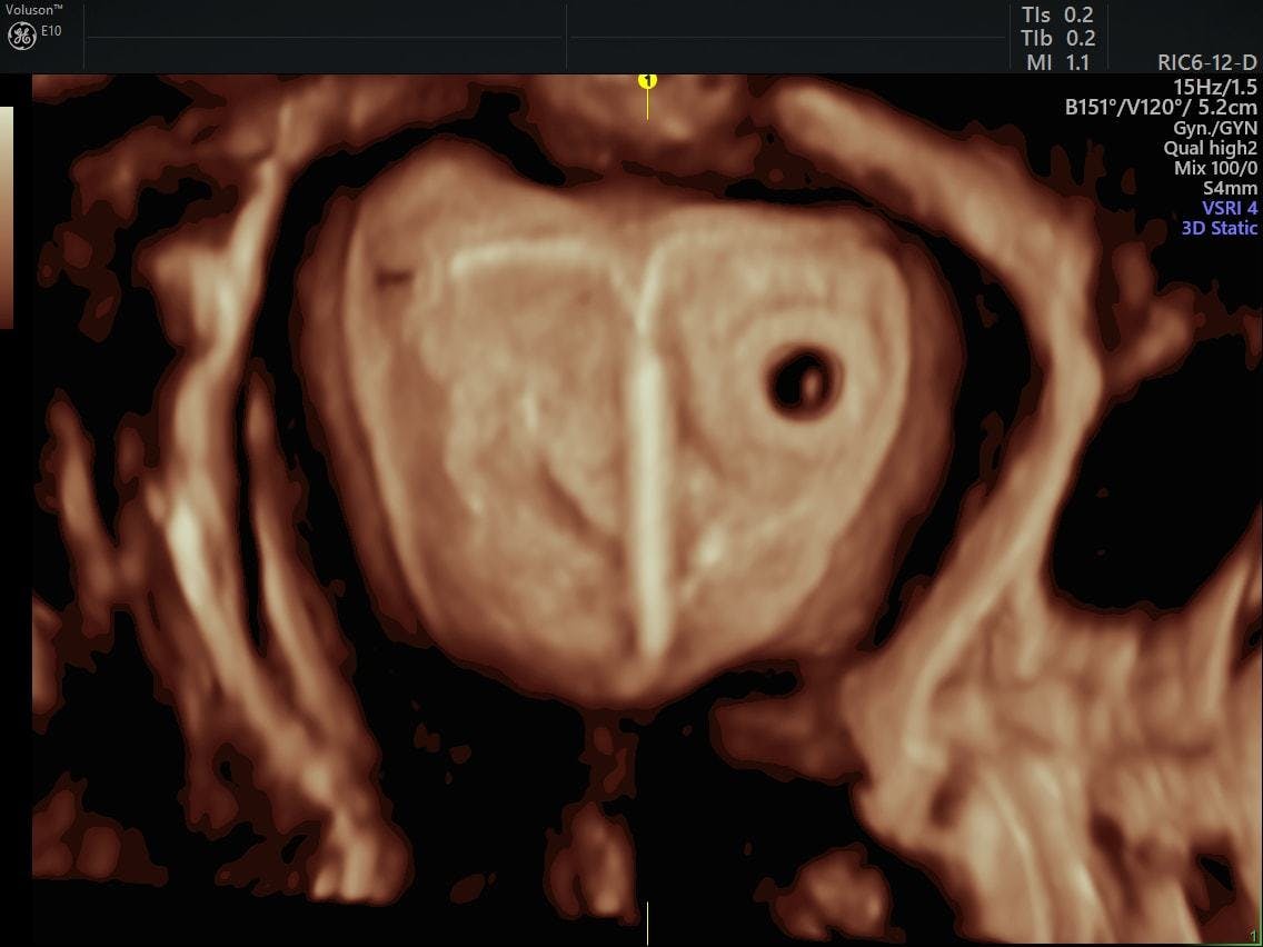
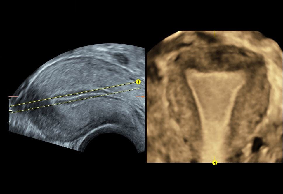
In this ultrasound the fetuss neck thickness is measured. The best time to get the 3d ultrasound is between week 26 and week 30 of your pregnancy. Ive seen parents come in worried that something was wrong with their baby because the photo didnt look right if you do decide to get a keepsake ultrasound youre more likely to get the pretty images you see in magazines between 24 and 28 weeks. As a result the 3d ultrasound will only pick up images of the bones of the face.
During this time the ultrasound will bring some amazing pictures of the baby. A special transducer is gently inserted into the vagina to get a quick look at the uterus and ovaries. Ultrasound is usually painless. The baby is very active during this stage of the pregnancy.
Below is a breakdown on what to expect during an ultrasound depending on how many weeks pregnant you are. Before your baby is 26 weeks old in the womb there will be little fat under his skin. Some 3 d ultrasound photos also can look a little scary. Keep in mind that 3d and 4d ultrasounds are not typically used to diagnose problems with your baby.
Experts also discourage the use of any kinds of ultrasounds 2d doppler 3d and 4d for the purpose of creating a memento.

3d Ultrasound 4d Ultrasound Olympia Inside View Ultrasound
3d Ultrasound Services In Reno Women S Health Center Of Reno
3d Ultrasound Houston And 4d Ultrasound Houston Picture Perfect Imaging 3d 4d Ultrasound

We Got A 4d Ultrasound 28 Weeks Youtube

Preview Mobile 3d 4d Ultrasound Llc Home Facebook